
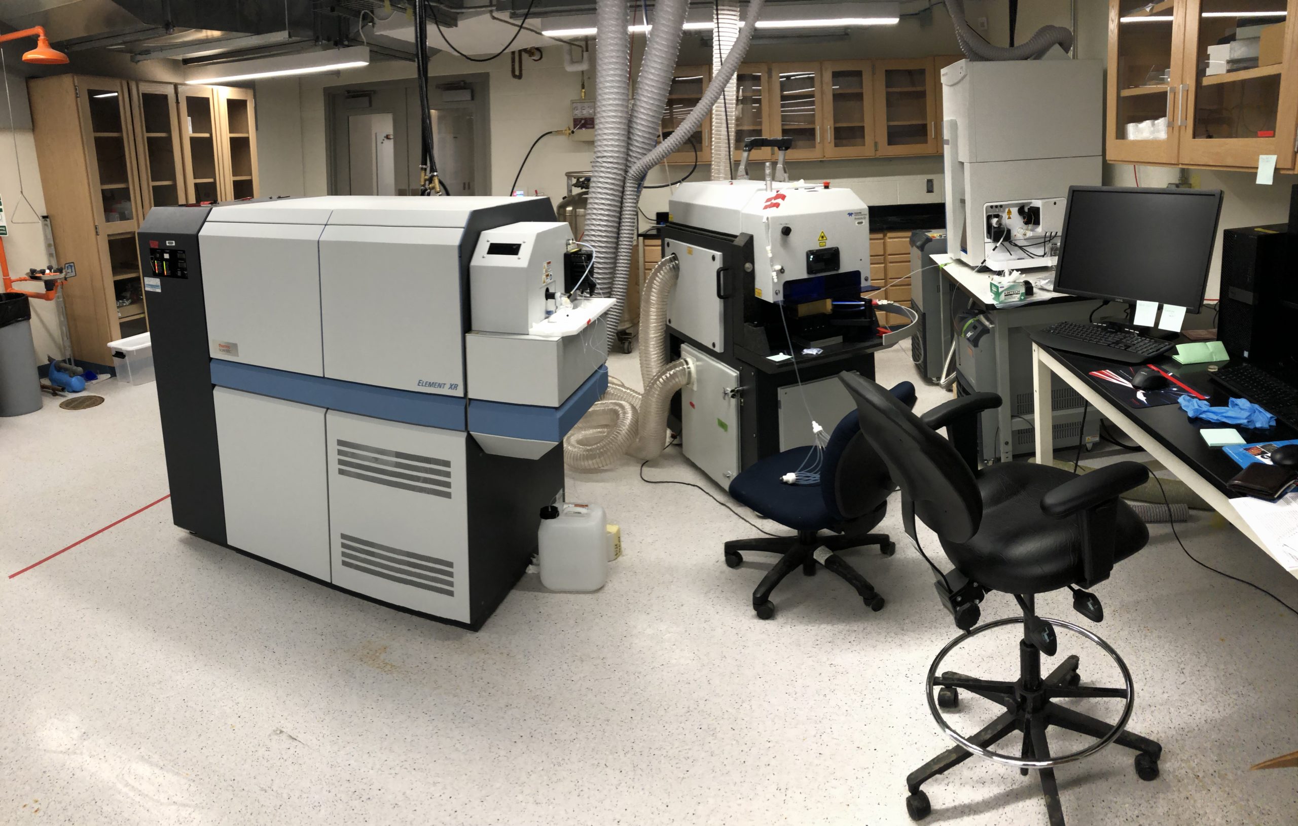
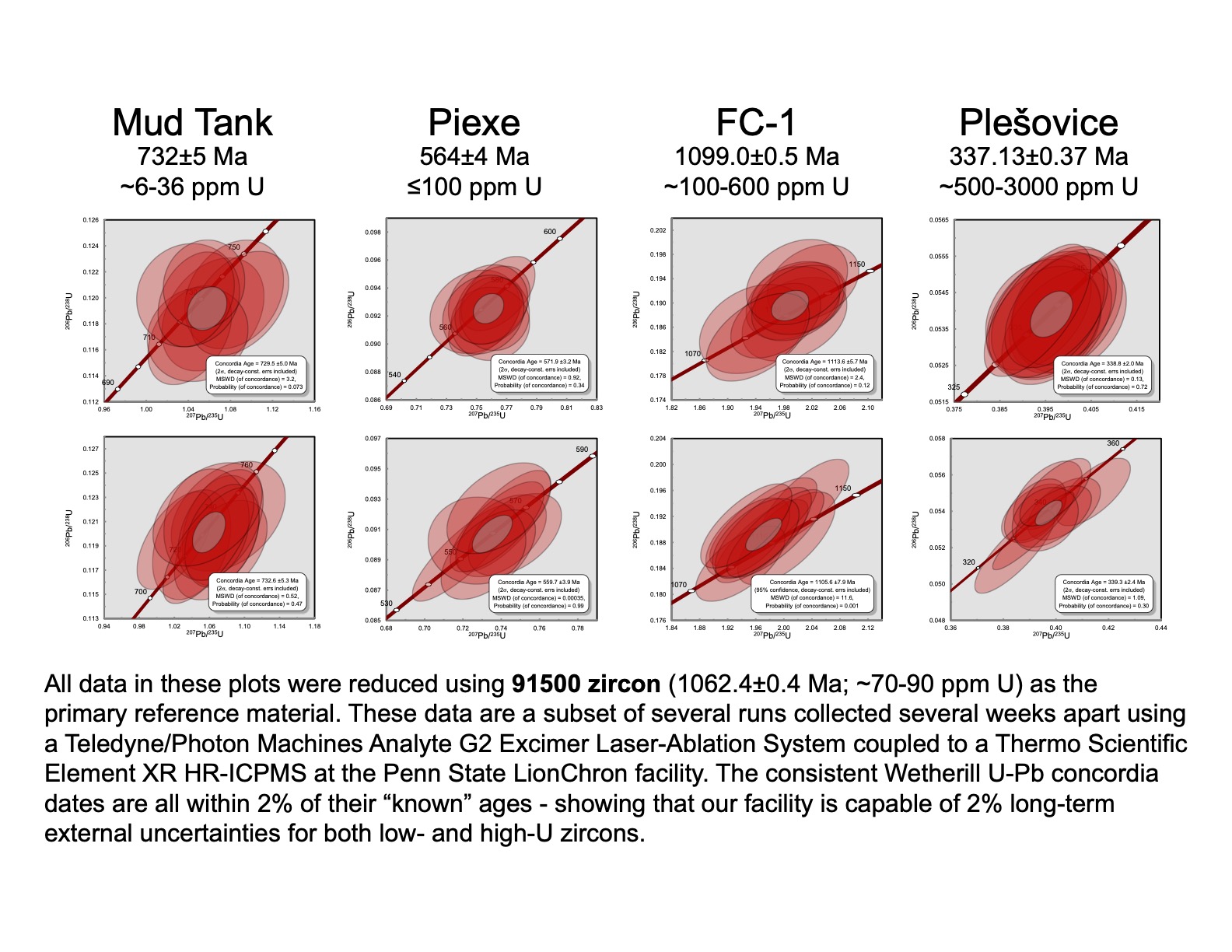
Laser-Ablation Split-Stream Facility
The LionChron facility hosts a Thermo Scientific Element XR HR-ICP-MS, a Thermo Scientific iCAP RQ-ICPMS, and a Teledyne/Photon Machines Analyte G2 Excimer Laser Ablation System (installed Dec 2020 – Jan 2021). The system is designed such that material ablated by the laser – either in thin section or epoxy grain mounts – can be sent to each mass spectrometer individually, or in tandem (split-stream). As of Spring 2021, we are routinely producing publication-quality U-Pb (zircon) + trace-element (zircon and other metamorphic phases) datasets, and are expanding to other dateable U-Pb phases (monazite, titanite, apatite).
Microscopy
The petrography laboratory at PSU is equipped with a suite of state-of-the-art optical microscopes to perform manual and automated characterization of rock sample petrography. These include: i) a Zeiss AxioImager A2.m petrographic microscope with Zeiss digital camera system (installed summer 2017); ii) a Zeiss Axioscope A1 routine Pol scope with digital imaging system (installed Feb 2020), and iii) a Zeiss AxioScan.Z1 fully-automated slide scanner, capable of digitizing standard and large format thin sections under PPL, XPL and epifluorescence at <10 μm resolution (installed Feb 2020).
Sample Preparation Facilities
PSU has rock preparation facilities that include (1) jaw crushers, pulverizers, and puck mills for breaking down xenoliths to the size fractions appropriate for mineral separation; (2) rock saws and lapidary equipment for sectioning and polishing samples for microscopy; and (3) mineral separation equipment, including heavy liquids separation funnels and accessories, a Frantz magnetic separator, and binocular picking microscopes. Additionally, the PSU Materials Characterization Laboratory hosts a general-purpose sample preparation lab with C- and Ir-coating equipment for preparing high-vacuum electron microscopy samples.
Computational Laboratory
[Coming soon]
SEM and EPMA
The PSU Materials Characterization Lab at the Mineral Research Institute operates three Scanning Electron Microscopes: i) a FEI NanoSEM 630 FESEM with 1.7 nm resolution, iii) an Apreo S SEM with a Symmetry EBSD detector (Thermo-Scientific), and iii) a Varios G4 SEM with an Oxford Nordlys Max2 EBSD detector. The PSU MCL also houses a Cameca SX5 Electron Probe Micro-Analyzer (EPMA) with simultaneous X-Ray, SEM and BSE imaging capability, three high sensitivity WD spectrometers, two four-crystal high resolution spectrometers and an LaB6 electron source. Each of these instruments is operated by a full-time PSU technician supported through the MCL.
TIMS
We operate a ThermoScientific TRITON Plus multi-collector thermal ionization mass spectrometer (TIMS). The instrument was installed in summer 2015 and is currently in use for high-precision Rb-Sr geochronology of metamorphic mineral phases and deformation fabrics. Microsampling of key phase assemblages is performed using a New Wave Micromill. The TRITON's detector block configuration includes an extra large L5 Faraday, ideally suited for 40Ca collection. We are currently measuring Sr to sub-permille precision and plan to establish Sm-Nd geochronology and Ca isotope measurements in the near future. The TIMS laboratory is a part of the Laboratory for Isotopes and Metals in the Environment (LIME).